Anatomy Pictures Of Lower Back And Hip : Pin On Health. Understanding the anatomy of your lower spine can help you communicate more effectively with the medical professionals who treat your lower back pain. This article looks at the anatomy of the back, including bones, muscles, and nerves. The anatomical areas found on the upper limb can serve as key landmarks to help us find important anatomical structures such as finding one of the superficial veins: Browse 4,815 hip anatomy stock photos and images available, or search for hip replacement or knee anatomy to find more great stock photos and pictures. The hip joint is a ball and socket synovial type joint between the.
Anatomy pictures of lower back and hip / sacroiliac si joint pain understanding causes symptoms treatment. Understanding lower back anatomy is key to understanding the root of lower back and hip pain. The muscles of the lower back help stabilize, rotate, flex, and extend the spinal column, which is a bony tower of 24 vertebrae that gives the body structure and houses the spinal cord. This article looks at the anatomy of the back, including bones, muscles, and nerves. This nerve runs from the lumbar plexus along the psoas major past the inguinal ligament to enter the femoral triangle.
Lower Back Pain Types Symptoms Treatment from cdn.sanity.io The lumbar region of the spine, more commonly known as the lower back, is situated between the thoracic, or chest, region of the spine, and the sacrum. To put it plainly, sometimes hip pain comes from the hip, but a lot of times hip pain comes from the back. Low back muscle spasming is common because lumbar extensor muscles must contract eccentrically. Anatomy pictures of lower back and hip : Possible causes of lower back and hip pain include sprains, strains, and a herniated disk. The vertebral column of the lower back includes the five lumbar vertebrae, the sacrum, and the coccyx. The hip joint is the uppermost part of the leg where the head of the thigh bone (femur) fits into the socket of the pelvis. They provide a great deal of strength to modulate powerful forces between the upper and lower body.
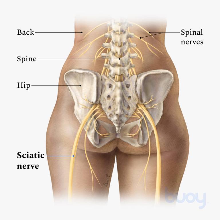
The sacrum is the bottom part of the spine, which connects to the hip bones.
When most people mention their back, what they are actually referring to is their spine. Anatomy pictures of lower back and hip. Karir para artis march 20, 2021 groin, inguinal region and the fascia / aponeurosis: Picture tests in practical anatomy. The different anatomical areas of the gluteal region: The lumbar region of the spine, more commonly known as the lower back, is situated between the thoracic, or chest, region of the spine, and the sacrum. It allows for complete rotations of the hip and is also. Hold each of these lower back and hip stretches for at least 15 to 30 seconds, and repeat several times on each side. Make sure you're stretching to the point of tension, not pain; When a person experiences lower back and hip pain simultaneously, there may be an underlying injury or medical. The muscles of the lower back help stabilize, rotate, flex, and extend the spinal column, which is a bony tower of 24 vertebrae that gives the body structure and houses the spinal cord. As well as some basic images of disc pathology and stylised facet joint motion. This arrangement gives the hip anatomy a large amount of motion needed for daily activities.
This nerve runs along the. The lumbar region of the spine, more commonly known as the lower back, is situated between the thoracic, or chest, region of the spine, and the sacrum. The anatomical areas found on the upper limb can serve as key landmarks to help us find important anatomical structures such as finding one of the superficial veins: Low back muscle spasming is common because lumbar extensor muscles must contract eccentrically. Anatomy pictures of lower back and hip :
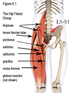
Tight Hip Flexors Causing Back Hip Pain Low Back Pain Program from lowbackpainprogram.com It has branches that innervate the anterior thigh muscles and the hip joint. Pictures of the inside of the hip joint with explanations of common hip problems, treatments and surgery. Make sure you're stretching to the point of tension, not pain; Anatomy of the lower extremity ii. The lumbar region of the spine, more commonly known as the lower back, is situated between the thoracic, or chest, region of the spine, and the sacrum. Browse 221 lower back skeleton stock photos and images available, or start a new search to explore more stock photos and images. Anatomy pictures of lower back and hip : A basic understanding of the anatomy of your lower back can help you identify and differentiate a problem that commonly.
Muscles of the lower back and hip diagram, human muscles, muscles of the lower back and hip diagram.
Anatomy pictures of lower back and hip : Understanding lower back anatomy is key to understanding the root of lower back and hip pain. See more ideas about anatomy, thoracic, basic image. The different anatomical areas of the gluteal region: Bones of the pelvis and lower back. Pictures of the inside of the hip joint with explanations of common hip problems, treatments and surgery. To put it plainly, sometimes hip pain comes from the hip, but a lot of times hip pain comes from the back. Anatomy pictures of lower back and hip / sacroiliac si joint pain understanding causes symptoms treatment. When something injures or puts pressure on the sciatic nerve, it can cause pain in the lower back that spreads to the hip, buttocks, and leg. As well as some basic images of disc pathology and stylised facet joint motion. Learn about anatomy lower limb with free interactive flashcards. This nerve runs along the. When a person experiences lower back and hip pain simultaneously, there may be an underlying injury or medical.
A basic understanding of the anatomy of your lower back can help you identify and differentiate a problem that commonly. By dr arun pal singh. When a person experiences lower back and hip pain simultaneously, there may be an underlying injury or medical. The different anatomical areas of the gluteal region: This arrangement gives the hip anatomy a large amount of motion needed for daily activities.
Pin On Anatomia from i.pinimg.com See more ideas about anatomy, thoracic, basic image. Up to 90% of people recover from sciatica without surgery. When something injures or puts pressure on the sciatic nerve, it can cause pain in the lower back that spreads to the hip, buttocks, and leg. It allows for complete rotations of the hip and is also. A basic understanding of the anatomy of your lower back can help you identify and differentiate a problem that commonly. The anatomy of the hip and back is comprised of numerous parts that can be injured or wear out, and many problems that occur in this area can display the exact same symptoms or pathology. The vertebral column consists of 33 vertebrae which can be split up into 5 continuous sections. Hip pain may be due to a variety of common causes including fractures, sprains, strains, arthritis, and bursitis.
The vertebral column of the lower back includes the five lumbar vertebrae, the sacrum, and the coccyx.
To put it plainly, sometimes hip pain comes from the hip, but a lot of times hip pain comes from the back. Normally, a smooth cushion of shiny white hyaline (or articular) cartilage about 1/4 inch thick covers the femoral head and the acetabulum.the articular cartilage is kept slick by fluid made in the synovial membrane (joint lining). The bones of the pelvis and lower back work together to support the body's weight, anchor the abdominal and hip muscles, and protect the delicate vital organs of the vertebral and abdominopelvic cavities. It has branches that innervate the anterior thigh muscles and the hip joint. When most people mention their back, what they are actually referring to is their spine. Anatomy pictures of lower back and hip. The hip joint is the uppermost part of the leg where the head of the thigh bone (femur) fits into the socket of the pelvis. The vertebral column of the lower back includes the five lumbar vertebrae, the sacrum, and the coccyx. The hip joint is a ball and socket synovial type joint between the. They provide a great deal of strength to modulate powerful forces between the upper and lower body. Hip pain may result from inflammation, degeneration, or injury to structures and tissues within the hip joint. Make sure you're stretching to the point of tension, not pain; Pictures of the inside of the hip joint with explanations of common hip problems, treatments and surgery.
Share :
Post a Comment
for "Anatomy Pictures Of Lower Back And Hip : Pin On Health"


Post a Comment for "Anatomy Pictures Of Lower Back And Hip : Pin On Health"